

Zero Lab Treatment Information
Not every brown patch is melasma
Hello, I am Dr. Dong Won Kim, Medical Director of Zero Lab Clinic, one minute from Hongdae (Hongik Univ.) Station Exit 1.
Of the people who come in asking us to "get rid of this melasma," only about half actually have melasma. The rest have freckles, or solar lentigines, or marks left by acne, or — rarely — a nevus of Ota they were born with. On the surface they are all brown patches, so we do not call it by eye alone. These four differ in the depth the pigment sits at, in why they form, and in how they respond to a laser.
Today I will go through how we tell brown patches on the face apart, and why melasma in particular is so difficult, with the research behind it.
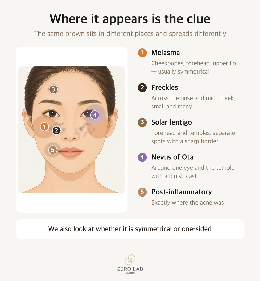
The model's face in the first image is an AI-generated illustration. It is not a real patient and not a before-and-after photograph.
The same brown, different roots
Let me separate the four first. Where it appears and the age it starts already narrow things down a great deal.
| Where it appears | When it starts | Depth the pigment sits at | |
|---|---|---|---|
| Melasma | Cheekbones, forehead, upper lip — usually symmetrical on both sides | Late twenties onward, after pregnancy or hormonal change | Spans epidermis and dermis |
| Freckles | Across the nose and mid-cheek, small and many | Young age, darkens with sun | Epidermis |
| Solar lentigo | Temples, forehead, backs of the hands — separate spots with a sharp border | Accumulates from middle age | Epidermis (often overlapping other lesions) |
| Post-inflammatory hyperpigmentation | Exactly where the acne or wound was | After the inflammation settles | Epidermis or dermis |
| Nevus of Ota | Around one eye and the temple, with a bluish cast | Just after birth, or at puberty | Dermis |
In the clinic we add a Wood's lamp and magnified examination to this. Pigment that sits only in the epidermis shows a sharper border under a different light; pigment in the dermis does not.
Symmetry is another important clue. Melasma often appears in a similar shape on both cheekbones, while a nevus of Ota is usually on one side. Post-inflammatory hyperpigmentation has nothing to do with symmetry — it simply follows wherever the inflammation was.
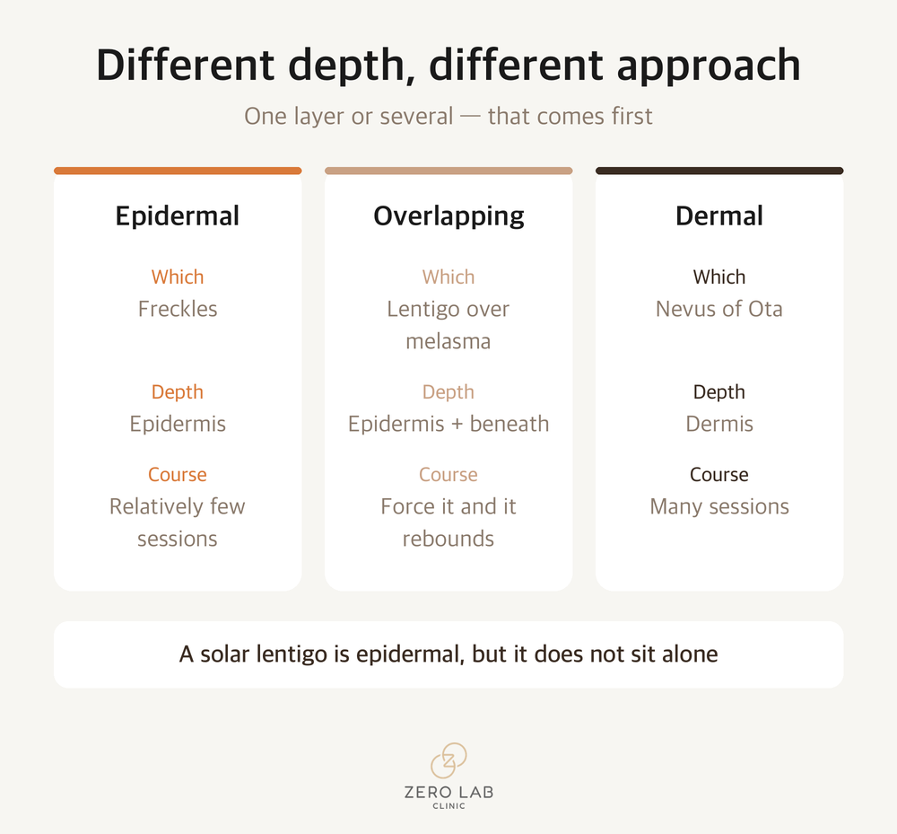
This distinction matters because the depth determines which devices can be used and how long it will take. Epidermal pigment tends to clear in relatively few sessions; dermal pigment takes far more. And as I will explain later, melasma has a character different from either of those.
Where is pigment made, and where does it go?
Since we keep coming back to depth, let me set out how pigment is made in the first place and where it goes. Once you know this, everything after it becomes much simpler.
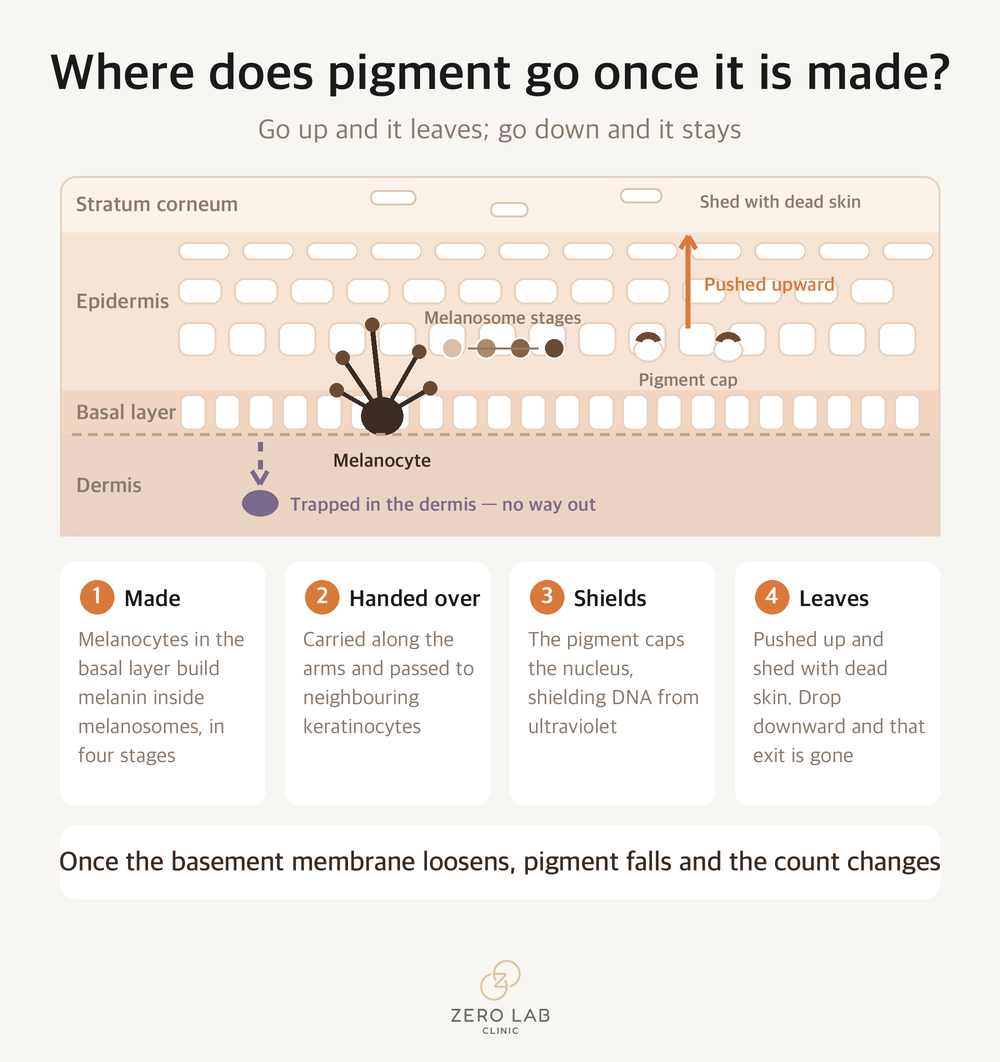
The cell that makes pigment is the melanocyte. It sits in the basal layer at the very bottom of the epidermis, and it cannot be seen with the naked eye. Even on a biopsy it takes a special stain to count them. And there is one thing worth noting here: in melasma, the number of these cells is usually normal. The colour does not deepen because there are more of them — they do more of what they already do.
This cell extends several branching arms, like an octopus. Melanin is made in stages inside a sac called a melanosome, four stages in all, and once finished it is carried along the cell's skeleton to the tips of those arms and handed over to the neighbouring keratinocytes. The pigment that crosses over settles like a cap over the keratinocyte's nucleus[14]. That is its original job — shielding the genetic material from ultraviolet light. Pigment is something the skin makes to protect itself, not the result of something breaking.
There is one more thing here. Melanocytes are not the only cells telling pigment to be made. Small vesicles released by keratinocytes have been shown to make melanocyte arms grow and to promote melanosome maturation and transfer[15]. Pigment is not the work of one cell but something the surrounding cells produce together.
Normally this pigment is pushed upward along with the keratinocytes and shed together with the dead skin cells. The epidermis turning over carries the pigment out with it. That is why epidermal pigment tends to clear in relatively few sessions. Push it up and there is an exit.
The problem is that in a diseased state a second route opens. When the basement membrane between epidermis and dermis loosens, or inflammation settles in that layer, melanin falls downward instead of moving up. Macrophages in the dermis collect the fallen pigment. Pigment that has dropped this far cannot leave with the dead skin cells.
So when we look at pigment, depth is the first thing we work out. Pigment in the epidermis has turnover as an exit; pigment that has dropped into the dermis does not. That is where the difference in the number of sessions begins, and where some of these conditions become things you manage rather than remove.
Why we do not resurface a solar lentigo away
In the table I placed solar lentigo on the epidermal side. That is correct. Solar lentigo is, histologically, an epidermal lesion. More melanin in the basal layer and elongated rete ridges — that is the definition.
The problem is that a solar lentigo rarely sits alone on a face. In practice the layers overlap. A lentigo sits on top of ground already laid with melasma, or on a dermis already changed by photoaging. What you see from outside is a single brown patch, but something else is there underneath it.
There is another way they overlap: when inflammation settles onto a lentigo. A Japanese study that followed 17 cases of lichen planus-like keratosis with dermoscopy over a year set out five stages of evolution, and the first stage is a pre-existing solar lentigo. When inflammation attaches to it, the pigment that was in the epidermis drops into the dermis, and pigment caught by dermal macrophages remains as blue-grey dots. Those dots take one to two years to disappear[13]. It began as an epidermal lesion and ended as dermal pigment.
That is why a plan built on removing only the epidermis does not fit well. The brown on the surface comes away, but what is underneath stays, and going deeper to clear the rest is where you start losing things.
The research reads the same way. There is a split-face study in 22 Asian patients using a low-fluence 1,064 nm Q-switched laser — the treatment usually called toning— for ten sessions. Small solar lentigines cleared by more than half in 62.5% of cases, but in larger solar lentigines the erythema index, vascularity and mast cell activity did not improve. The authors conclude that lesion thickness, vascularity and mast cell activity can predict the treatment response[10]. If the pigment were only in the epidermis, there would be no reason for thickness or vascularity to decide the outcome. It means there is something more underneath.
The approach used to be different. The epidermis was removed wholesale with an ablative laser, and toning was often layered on top of that. It was a way of forcing out the pigment that remained.
We do not do much of that now. A review of how melasma treatment has evolved records that high-energy pigment lasers, ablative lasers and fractional lasers left an unacceptably high rate of post-inflammatory hyper- and hypopigmentation, and rebound melasma[11]. A review of combination therapy with energy-based devices is more explicit. It states that combining ablative techniques with a Q-switched laser is not recommended, because of the high risk of permanent adverse effects such as guttate hypopigmentation and of making melasma worse[12].
On our skin the problem is larger. A face with a solar lentigo and nothing else is rare. Melasma is often lying underneath as well. Go in hard aiming at the lentigo and that stimulus wakes the melasma beside it. Colour that has come back as a rebound is harder to handle than it was at the start.
So we do not plan to clear a solar lentigo in one go either. We look first at what is lying underneath, and if the layers overlap we spread the work over more sessions. The colour comes away slowly, but we take the side that leaves nothing irreversible behind.
Melasma is not a problem of pigment alone
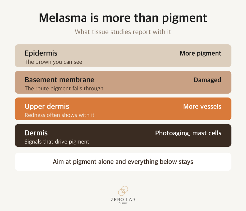
If you understand melasma only as "more melanin," treatment does not go well. What tissue studies say consistently is that in melasma-affected skin, several layers have changed together — not only the pigment cells.
A review of melasma pathology in an international journal reports that melasma lesions show increased epidermal pigment together with basement membrane damage, dermal photoaging (solar elastosis), increased vascularity and increased mast cells[1]. Melasma looks like an epidermal condition, but dermal factors play a central role.
A 2025 review adds another layer. Dermal fibroblasts placed under ultraviolet stress shift into a senescent state and keep secreting signalling molecules that stimulate pigment production, and immune cell infiltration — T cells, macrophages, mast cells — is seen within the lesion[2]. Another 2025 review likewise concludes that melasma should be seen not as a simple pigment disorder but as a chronic inflammatory microenvironment in which epidermis and dermis act on each other[3]. How much sunscreen you actually need to apply is covered separately in Sunscreen: reapplying matters more than the number.
To sum up: in melasma the brown you see is the outcome, and underneath it lies a state where the basement membrane has loosened, vessels have increased and inflammatory signals are circulating. Aim at the pigment alone and the cause stays exactly where it was.
Ultraviolet is not the only thing involved in melasma
Sun protection is the basis of managing melasma. But there is one more thing.
The 2025 review cited above treats visible light and hormonal fluctuationas separate items in the pathophysiology of melasma[2]. Visible light has a longer wavelength than ultraviolet, passes through window glass, and comes from indoor lighting and screens as well. The problem is that what SPF and PA measure on an ordinary sunscreen is ultraviolet. Visible light is not filtered by those numbers.
Let me bring in a familiar distinction here: chemical and mineral sunscreen. Chemical (organic) sunscreencontains ingredients that absorb ultraviolet and dissipate it, while mineral (inorganic) sunscreenworks by having particles such as zinc oxide and titanium dioxide bounce ultraviolet away.
But what both of them block is ultraviolet. Neither blocks much visible light at all. Chemical or mineral, this part is the same.
What blocks visible light is colour. Only when a coloured mineral ingredient such as iron oxide is included does the visible-light range get covered. In cosmetics this is called a tinted formulation — one that carries a skin tone. That is why we suggest a tinted product to people with melasma. It leaves a slight colour on the face, and that colour is doing work.
That said, this is not to say "use a tinted product and melasma will improve." It is closer to a piece that explains why melasma darkens again every summer. Some people tell us the colour rises even though they stayed indoors, and that makes sense once you account for visible light.
How to apply sunscreen is covered in a separate article. The short version is that the amount comes before the SPF number, and if you have melasma it is worth reading alongside this one.
Marks left by acne are not the same as melasma
A good proportion of the people who come in about pigment actually have post-inflammatory hyperpigmentation — brown left behind where acne or a wound settled.
Telling these two apart matters because they go in different directions over time. Post-inflammatory hyperpigmentation moves towards fading once the inflammation driving it stops, even if it takes a while. Melasma does not. The recurrence rate above shows that difference[5].
So when acne marks are mixed in, the order matters. If you aim at pigment while inflammation is still rising, the stimulus from the treatment can come back as fresh pigmentation. We settle the inflammation first, then look at the pigment.
We have written separately about acne scar care; pigment and scarring often appear together, so it is worth reading both.
This is why melasma is "managed," not "erased"
So that this does not sound abstract, let me show it in numbers.
In a retrospective study of 40 melasma patients in Korea given the same toning, the melasma severity score (mMASI) fell by an average of 54.23% after a median of ten sessions. In the same study, however, two patients developed mottled hypopigmentation and rebound pigmentation[4].
What deserves more attention is the long-term follow-up. In a randomised split-face study comparing a 755 nm picosecond laser with a 1,064 nm Q-switched laser, among the 17 patients followed for two years, 10 (58.82%) reported recurrence or worsening. There was no difference in recurrence between the two lasers[5].
In other words, even with good treatment a substantial proportion see it come back. That is not because the treatment was wrong; it is the nature of melasma as a condition. So when we are asked in consultation "how many sessions until it is gone," we do not answer with a number of sessions. We answer with a period of time that includes sun protection and maintenance.
What happens when toning is repeated
Toning is widely used for melasma because there genuinely is short-term improvement. But increasing the number of sessions does not translate directly into results.
In a study of 19 patients with mixed-type melasma given six sessions of a low-fluence Q-switched laser on one side of the face, mottled hypopigmentation with indistinct borders appeared in 21.05% by the sixth session[6]. The pigment fades and then leaves what look like white dots instead. Once this appears it is awkward to reverse.
The rebound pigmentation in the Korean study above belongs to the same picture[4]. When the stimulus is repeated, pigment cells turn back towards being activated again.
Changing the interval matters more than the number of sessions
Does that mean toning should not be done? No. It is also true that in the Korean study the melasma severity score fell by an average of 54.23% after a median of ten sessions[4]. The problem is carrying on at the same interval.
When we set out a course of pigment treatment, we explain it in two stages.
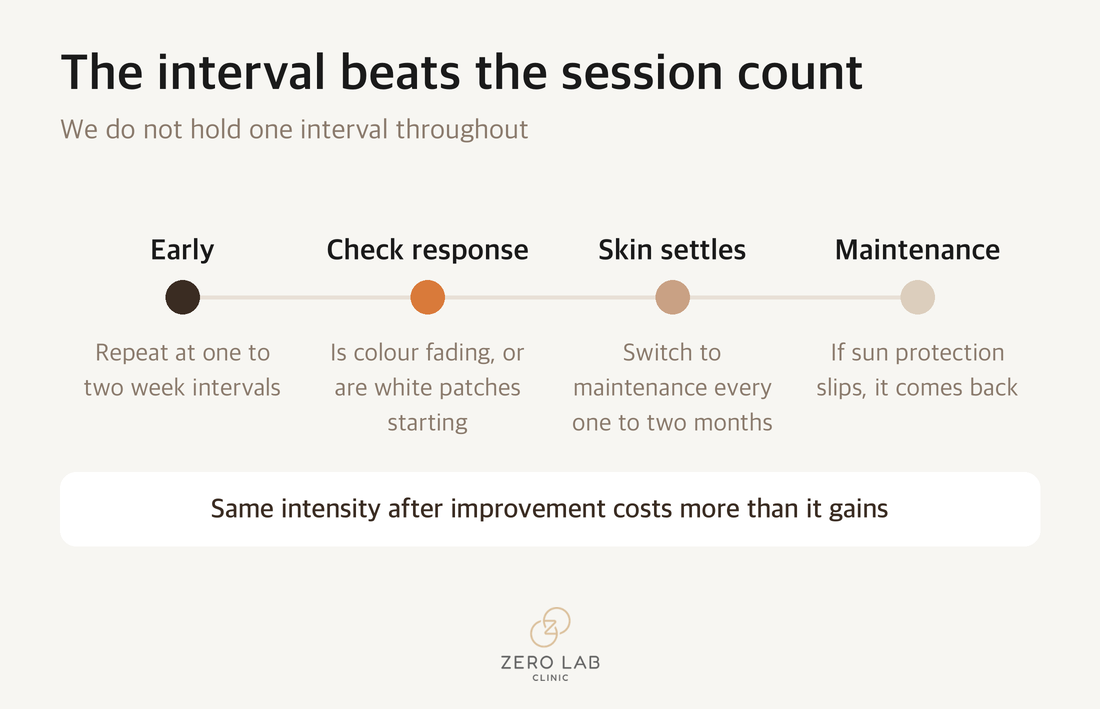
① Early — one to two week intervals
At the start we keep the intervals short. Pigment does not come away in one go; it fades a little at a time over several sessions, so some repetition is needed early on. The studies landing around ten sessions were describing this phase.
② Once the skin settles — switching to maintenance at one to two month intervals
Once the colour has visibly faded and the skin has stabilised, we do not hold the same interval. We widen it. We move to once every one to two months and maintain from there.
There are two reasons this switch matters.
One is the adverse effects above. The report of hypopigmentation in 21.05% by the sixth session[6] and the report of rebound pigmentation[4] both belong to what happens when stimulus keeps stacking up at short intervals . Carrying on at the same intensity after things have improved means losing more than you gain.
The other is recurrence. That 58.82% reported recurrence or worsening at two years[5] means, turned around, that what decides the outcome is not when treatment ends but how it is maintained. Melasma is not a condition you settle once and finish, so a stretch of holding on to the faded state is needed in its own right.
So what we watch is not the session count but this.
- Colour fading and white patches starting to appear are not the same thing. We work along that boundary
- When the response improves we widen the interval rather than using up the remaining sessions
- If sun protection slips, it comes back during the maintenance phase
Nevus of Ota is a different story
Talking only about melasma can make every pigment condition sound difficult, and that is not the case. A nevus of Ota has its pigment in the dermis so it takes many sessions, but it responds properly.
In a study dividing 81 nevus of Ota patients into two arms, the low-fluence 755 nm Q-switched alexandrite laser arm scored higher for improvement, and post-inflammatory hyperpigmentation after treatment was lower at 4.8% against 25.6%[7]. That turning up the energy is not always better connects with what I said about melasma.
When treatment starts also affects the outcome. In a study following 84 nevus of Ota patients over ten years, starting treatment before the age of five took an average of 2, 4 and 7 sessions to reach 25%, 50% and 75% pigment reduction, while starting after five took 3, 7 and 11 sessions. The risk of post-inflammatory hyperpigmentation was also significantly lower in the group that started early[8].
That said, this study includes paediatric patients, and it does not mean treatment fails in adults. Read it as meaning it takes more sessions.
Some brown patches need to be identified first
This is the part I most want to say.
A study analysing 1,197 atypical pigmented facial lesions reports that benign lesions such as lentigines, actinic keratosis and seborrhoeic keratosis and malignant lentigo maligna share overlapping clinical and dermoscopic features. The nose in particular was less clearly separable, as features of both benign and malignant lesions could appear together there[9].
So for a patch that appeared suddenly, or that is growing on one side only, or whose colour is uneven, or whose border is spreading, we do not start with a laser. Establishing what it is comes first. A pigment laser lightens colour, so using one on a lesion that needs to be identified can make the judgement harder.
What we check in consultation
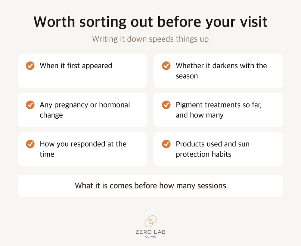
When you come in about pigment, we ask the following first. Having it ready makes the consultation much faster.
1) When it appeared, and whether it darkens with the season
2) Whether there has been pregnancy, breastfeeding or any hormonal change
3) The type and number of pigment treatments you have had, and how you responded
4) The products you are using and your sun protection habits
5) Whether it is a site where you had acne or inflammation
The question I am asked most is how many sessions it takes to be gone. What I say is that before that, we have to work out what it is. Melasma is a condition you manage, so it is better to include the maintenance stage when the plan is first drawn up. As for what to sort out first, bringing the five points above usually settles the direction.
Pigment left after acne runs a different course, so we covered that in the scarring article.
Treatments covered in this article
Prices and booking
References
[1] Kwon SH, Hwang YJ, Lee SK, Park KC. Heterogeneous Pathology of Melasma and Its Clinical Implications. Int J Mol Sci. 2016;17(6):824. DOI: https://doi.org/10.3390/ijms17060824
[2] Ali L, Al Niaimi F. Pathogenesis of Melasma Explained. Int J Dermatol. 2025;64(7):1201-1212. DOI: https://doi.org/10.1111/ijd.17718
[3] Miao F, Wan J, Zhou Y, Shi Y. Unraveling Melasma: From Epidermal Pigmentation to Microenvironmental Dysregulation. Biology (Basel). 2025;14(10):1402. DOI: https://doi.org/10.3390/biology14101402
[4] Choi JE, Lee DW, Seo SH, Ahn HH, Kye YC. Low-fluence Q-switched Nd:YAG laser for the treatment of melasma in Asian patients. J Cosmet Dermatol. 2018;17(6):1053-1058. DOI: https://doi.org/10.1111/jocd.12760
[5] Zhou Y, Li Y, Hamblin MR, Wen X. Comparison of 755-nm picosecond alexandrite laser versus 1064-nm Q-switched Nd:YAG laser for melasma: A randomized, split-face controlled, 2-year follow-up study. Lasers Surg Med. 2024;56(3):263-269. DOI: https://doi.org/10.1002/lsm.23763
[6] Saleh F, Moftah NH, Abdel-Azim E, Gharieb MG. Q-switched Nd:YAG laser alone or with modified Jessner chemical peeling for treatment of mixed melasma in dark skin types. J Cosmet Dermatol. 2018;17(3):319-327. DOI: https://doi.org/10.1111/jocd.12444
[7] Wu X, Wang X, Shang Y, Xu H, Zhang Z. Beneficial Effects of Treatment With Low-Fluence 755-nm Q-Switched Alexandrite Laser for Nevus of Ota. Lasers Surg Med. 2021;53(10):1364-1369. DOI: https://doi.org/10.1002/lsm.23421
[8] Achavanuntakul P, Manuskiatti W, Wanitphakdeedecha R, Jantarakolica T. Early Treatment Initiation Improves Outcomes in Nevus of Ota: A 10-Year Retrospective Study. Am J Clin Dermatol. 2022;23(1):105-114. DOI: https://doi.org/10.1007/s40257-021-00637-0
[9] Tognetti L, Cartocci A, Cinotti E, et al. Dermoscopy of atypical pigmented lesions of the face: Variation according to facial areas. Exp Dermatol. 2023;32(12):2166-2172. DOI: https://doi.org/10.1111/exd.14941
[10] Kaminaka C, Furukawa F, Yamamoto Y. The Clinical and Histological Effect of a Low-Fluence Q-Switched 1,064-nm Neodymium:Yttrium-Aluminum-Garnet Laser for the Treatment of Melasma and Solar Lentigenes in Asians. Dermatol Surg. 2017;43(9):1120-1133. DOI: https://doi.org/10.1097/DSS.0000000000001120
[11] Kauvar ANB. The evolution of melasma therapy: targeting melanosomes using low-fluence Q-switched neodymium-doped yttrium aluminium garnet lasers. Semin Cutan Med Surg. 2012;31(2):126-32. DOI: https://doi.org/10.1016/j.sder.2012.02.002
[12] Iranmanesh B, Khalili M, Mohammadi S, Amiri R, Aflatoonian M. The efficacy of energy-based devices combination therapy for melasma. Dermatol Ther. 2021;34(3):e14927. DOI: https://doi.org/10.1111/dth.14927
[13] Watanabe S, Sawada M, Dekio I, Ishizaki S, Fujibayashi M, Tanaka M. Chronology of lichen planus-like keratosis features by dermoscopy: a summary of 17 cases. Dermatol Pract Concept. 2016;6(2):29-35. DOI: https://doi.org/10.5826/dpc.0602a06
[14] Fukuda M. Rab GTPases: Key players in melanosome biogenesis, transport, and transfer. Pigment Cell Melanoma Res. 2021;34(2):222-235. DOI: https://doi.org/10.1111/pcmr.12931
[15] Prospéri MT, Giordano C, Gomez-Duro M, et al. Extracellular vesicles released by keratinocytes regulate melanosome maturation, melanocyte dendricity, and pigment transfer. Proc Natl Acad Sci U S A. 2024;121(16):e2321323121. DOI: https://doi.org/10.1073/pnas.2321323121
The 15 papers above are indexed in PubMed. This article is general medical information; decisions about an individual's condition are made through consultation.
This article was written directly by Zero Lab Clinic to provide medical information. Results of any procedure vary with individual skin condition, and side effects are possible. Please decide on a procedure only after an in-person consultation with a doctor.